
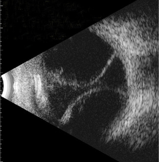
B-scan- things you must know about it
B-scan ultrasonography, also known as brightness scan, is a non-invasive diagnostic imaging technique used in ophthalmology to visualize the internal structures of the eye when the direct view is obscured especially when the media (like the cornea, lens, or vitreous) are opaque due to cataract, hemorrhage, or trauma.
Indications for B-scan
This test is particularly helpful when a routine fundus examination using ophthalmoscopy is not possible. Some common indications include:
- Dense cataract or vitreous hemorrhage
- Retinal detachment suspicion
- Ocular tumors (like choroidal melanoma)
- Intraocular foreign bodies
- Posterior scleritis
- Optic nerve head drusen
- Ocular trauma
Frequency
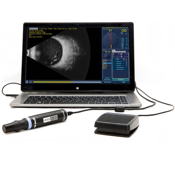
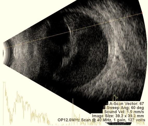
B-scan ultrasonography operates using high-frequency sound waves, typically in the range of 8 to 10 MHz, which provide sufficient resolution to visualize intraocular structures without penetrating beyond the eye. These ultrasound waves are emitted by a transducer probe that is placed either over the closed eyelid or directly on the anesthetized eye with the use of a coupling gel to ensure proper transmission. As the sound waves travel through the eye, they encounter various tissues that reflect the waves back at different intensities based on their density and composition. These returning echoes are captured by the transducer and converted into a two-dimensional, real-time cross-sectional image displayed on the screen. This allows clinicians to assess the anatomical relationships and detect abnormalities within the posterior segment of the eye, even when direct visualization is obstructed by opacities such as cataracts, corneal scars, or vitreous hemorrhage.
Gain
Gain in B-scan ultrasonography controls the sensitivity of the machine to returning echo signals, affecting the brightness of the image. High gain settings (90–100 dB) are useful at the beginning of the scan to detect low-reflective structures like vitreous hemorrhage or vitritis, but excessive gain can introduce noise, obscuring real anatomical details. Conversely, low gain settings (50–60 dB) are ideal for visualizing solid, highly reflective lesions such as tumors, as they reduce background noise and enhance structural clarity. A practical approach is to begin with high gain to localize abnormalities, then gradually decrease the gain for a sharper, more defined image.
Modes of B-scan
- Transverse scan: Probe placed horizontally across the eyelid. Good for peripheral retina and horizontal sections.
- Longitudinal scan: Probe placed vertically. Used for detailed views of anterior-posterior axis.
- Axial scan: Probe aligned along the visual axis. Best for optic nerve head and posterior pole visualization.
Structures to be seen in B-scan
- Anterior structures- though not ideal for cornea or anterior chamber evaluation, limited views may be possible.
- Lens- especially if it’s dislocated.
- Vitreous- can detect hemorrhage, inflammation (vitritis), or asteroid hyalosis.
- Retina- especially useful in cases of retinal detachment or tumors.
- Choroid- helps identify choroidal detachments, melanoma, or metastasis.
- Optic nerve head- useful in detecting optic nerve head drusen or swelling.
- Posterior sclera and orbit- detects posterior scleritis or orbital lesions.
Limitations for ocular structures
- Cornea
- Iris and anterior chamber angle
- Ciliary body (partial view possible in some cases)
- Retinal layers detail (OCT is more suitable here)
- Very small foreign bodies (if below resolution threshold)
Advantages
- Non-invasive and quick
- Portable and cost-effective
- Can be used bedside or in trauma settings
- Helps differentiate between retinal and choroidal detachments
Limitations for the machine
- Operator-dependent technique
- Cannot provide retinal layer detail like OCT
- Less accurate for anterior segment
- May miss subtle lesions in normal media
10 Eye Conditions Where B-Scan is Necessary for Diagnosis
Vitreous hemorrhage
In B-scan, vitreous hemorrhage appears as low-to-medium reflective, mobile dot-like or clump-like echoes within the vitreous cavity. These echoes move with eye movements and may obscure the view of the retina, especially in dense hemorrhage.
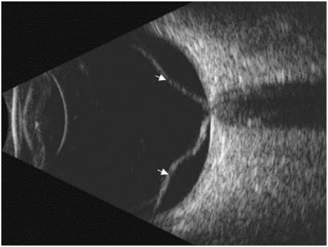
Retinal detachment
Retinal detachment on B-scan appears as a bright, V- or Y-shaped highly reflective membrane that moves independently with eye movement and remains tethered at the optic disc.
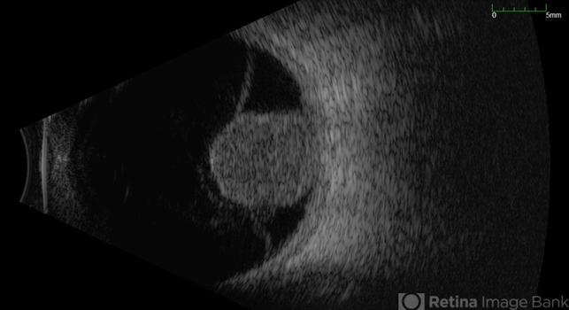
Choroidal melanoma
Choroidal melanoma appears on B-scan as a dome-shaped or mushroom-shaped, solid, highly reflective mass that shows internal low-to-medium echoes and may cause acoustic shadowing behind the lesion.
Posterior Vitreous Detachment (PVD)
Posterior Vitreous Detachment (PVD) on B-scan appears as a thin, mobile, low-reflective membrane that moves freely within the vitreous and is not attached to the optic disc, helping differentiate it from retinal detachment.

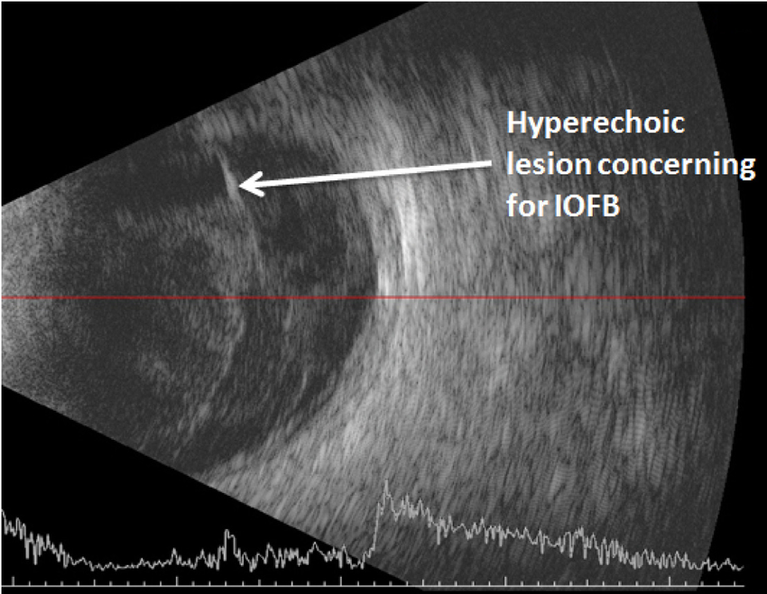
Intraocular foreign body (IOFB)
An intraocular foreign body (IOFB) appears on B-scan as a highly reflective echo with strong acoustic shadowing behind it; it remains stationary and may vary in size and shape depending on the material (e.g., metal, glass).
Endophthalmitis
Endophthalmitis on B-scan shows low-to-medium reflective echoes scattered throughout the vitreous, often appearing as dense, mobile opacities. The vitreous may look hazy or filled with debris, and in severe cases, the retinal details may be obscured.
Toxic Posterior Segment Syndrome (TPSS)
Toxic Posterior Segment Syndrome (TPSS) on B-scan may show diffuse, low-to-medium reflective echoes in the vitreous cavity, resembling vitritis. The retina typically remains attached, and there’s no evidence of retinal detachment. The findings are often subtle and must be correlated with recent intraocular surgery or exposure to toxic agents.
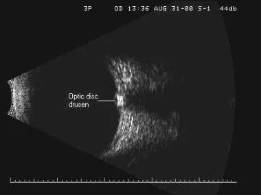
Optic disc drusen
Optic disc drusen on B-scan appear as small, highly reflective calcified spots within the optic nerve head, often with posterior acoustic shadowing. They are typically stationary and help differentiate drusen from true optic disc edema.
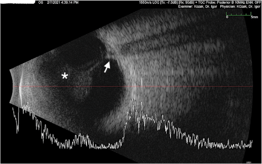
Posterior scleritis
Posterior scleritis on B-scan shows a characteristic “T-sign,” formed by fluid in the sub-Tenon’s space creating a hypoechoic area that joins the optic nerve shadow. The sclera may appear thickened, and there may be associated retinal or choroidal folds.

Suprachoroidal hemorrhage
Suprachoroidal hemorrhage or detachment appears on B-scan as smooth, dome-shaped, highly reflective choroidal elevations. In severe cases, the elevated choroids may touch, forming a “kissing” sign. If hemorrhagic, low-to-medium internal echoes may be seen in the suprachoroidal space.
Founder of EyesMatterMost- an optometry student who loves talking about eyes. I tend to cover topics related to optometry, ophthalmology, eye health, eyecare, eye cosmetics and everything in between. This website is a medium to educate my readers everything related to eyes.