Why ophthalomologists avoid cataract surgery in high myopia?
Have you ever wondered that why your ophthalmologist avoids and delays operating on your cataract if you use very high minus prescription ir have high myopia? This is because people with high myopia (pathological myopia) undergo structural changes in the eye leading to complicaitons in the retina.
High myopia and retinal pathologies
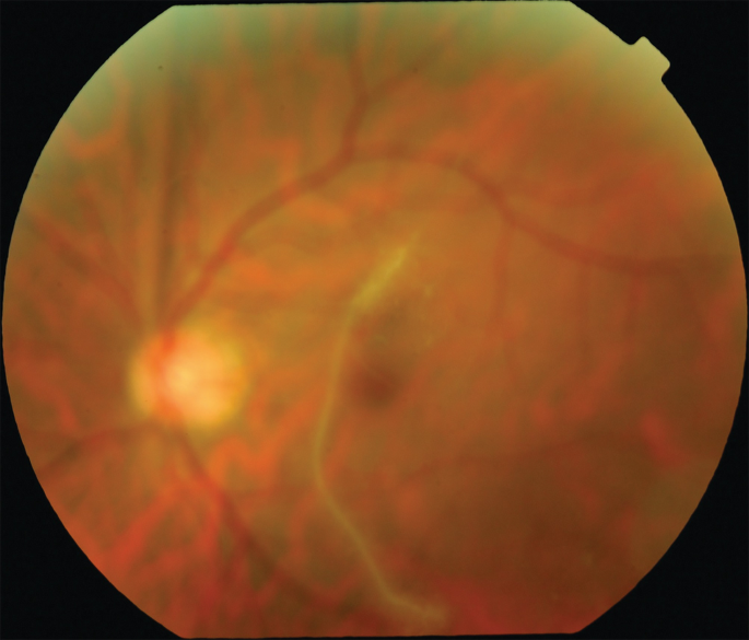
As the axial length of the eye continues to elongate, several ocular changes occur, including tilted optic discs, retinal thinning, and peripapillary atrophy.
The optic nerve head appears slanted or oblique because of the stretched posterior sclera and unequal growth of the globe.
The retina becomes progressively thinner, especially near the optic disc. This leads to PPA, where the tissue surrounding the optic disc degenerates. It appears as a grayish or whitish zone around the disc and indicates damage to the retinal pigment epithelium and choroid.
Increased risk of retinal detachment in pathological myopia
The structural fragility of a highly myopic eye makes it more prone to retinal breaks and detachments. Factors contributing to this include:
- Stretched and thin retina, which is more susceptible to tears.
- Posterior staphyloma, a bulging of the sclera at the back of the eye, which distorts the retinal architecture.
- Lattice degeneration, commonly seen in myopic eyes, which predisposes the retina to holes or tears.
Cataract Surgery: A careful decision in high myopia
Cataract surgery can increase the risk of retinal detachment in pathological myopia. This is primarily due to:
- Vitreous changes and zonular instability, which are more common in long axial length eyes.
- Sudden decompression of the posterior segment during surgery, which may lead to traction on the retina.
- Increased risk of posterior capsular rupture, leading to vitreous loss and retinal complications.
Posterior vitreous detachment (PVD)– blessing in disguise
Interestingly, if a patient already has a complete posterior vitreous detachment before undergoing cataract surgery, the risk of retinal detachment may be significantly lower. Why?
PVD eliminates vitreoretinal traction risk, which is a major mechanism leading to retinal tears.A detached vitreous means less pulling force on the thin retina during intraocular manipulation or postoperative vitreous shifts.
Founder of EyesMatterMost- an optometry student who loves talking about eyes. I tend to cover topics related to optometry, ophthalmology, eye health, eyecare, eye cosmetics and everything in between. This website is a medium to educate my readers everything related to eyes.