Aniseikonia- everything you need to know about
Aniseikonia is the perception of images of different sizes by each eye. The condition of perceiving different size of the images is associated with different refractive status of the eyes. When both the eyes have different refractive error, then this condition is called anisometropia.
How refractive changes take place
The changes in the refractive state can happen as a result of monocular changes in the axial length, amblyopia, curvature of cornea, retinal pathologies, lens, monocular ocular trauma, inflammation in either eye and strabismus, uni-ocular aphakia etc. These changes bring about variations in the refractive status in the eye that can result in anisometropia and ultimately aniseikonia if not treated properly.
Signs and symptoms
Our eyes are the extensions of our brain. Our brain deals with both of our eyes simultaneously. This state of unequal images can result in:
- Headaches
- Eye strains
- Nausea
- Difficulty in understanding distances
- Poor coordination
- Distortions of image
Causes
Variations in the refractive status of eyes can lead to changes in the size of images perceived by the visual areas of brain. The greater the differences in the refractive status of eye the greater is the aniseikonia.
Certain eye surgeries, such as cataract surgery or refractive surgeries like LASIK, can sometimes result in aniseikonia, particularly if there’s a disparity in the postoperative refractive error between the eyes.
Injuries to the eyes or head trauma can lead to structural changes that cause differences in the shape or size of the eyes, contributing to aniseikonia.
Anisometropia refers to a condition where there is a significant difference in the refractive error between the two eyes, which can lead to aniseikonia.
Also known as “lazy eye,” amblyopia can result in differences in visual acuity between the eyes, potentially causing aniseikonia.
How to diagnose aniseikonia
As an eyecare professional, we can diagnose aniseikonia by
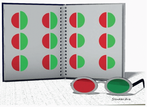
Eikonometer
It is the instrument with which the relative size of the images is calculated.
Comparison of image magnificaiton
It is also calculated by comparing the magnification of the images caused by the spectacles.
Major types
Follwing are one of the major types of aniseikonia
Optical aniseikonia
Optical aniseikonia refers to differences in image size or shape perceived by each eye due to variations in the optics of the eyes, particularly differences in refractive error or magnification. It typically arises from differences in the optical properties of the eyes, such as variations in the curvature of the cornea, the shape of the lens, or the axial length changes of the eye. These optical differences can lead to variations in the size, shape, or clarity of images projected onto the retina of each eye. Treatment may involve prescribing corrective lenses ( spectacles or contact lenses) or employing optical strategies to achieve more balanced visual perception.
Retinal aniseikonia
Retinal aniseikonia, on the other hand, involves differences in image size or shape perceived by each eye due to variations in the retinal image size or shape. In retinal aniseikonia there are variations in retinal size or shape, often associated with retinal disorders or structural abnormalities . Diseases like macular degenerations, retinal detachment, retinal scarring etc. can result in retinal aniseikonia. According to some researches, due to retinal pathologies there are mechanical distortions specifically micropsia in retinal aniseikonia.
Limitations to measuring aniseikonia
Subjective nature testing
It is often perceived subjectively by individuals, meaning that they may have difficulty accurately describing the differences in their visual perception. This subjective nature can make it challenging to quantify precise measurements through traditional methods.
Variations over menifestations
Aniseikonia can vary in degree and manifestation from person to person, as well as within the same individual over time. Even the differences in lighting conditions can influence the perceived magnitude of image.
Changing refractive status
The countinously changing refractive status also makes the previous readings invalid. It also puts barrier to measuring aniseikonia.
Lack of standard
There is a lack of widely accepted and standardized methods for measuring aniseikonia. There is no universally recognized gold standard for assessing and quantifying aniseikonia.
Binocular complexities
The complexities of binocular vision, including factors such as eye alignment, fusion, and stereopsis, can complicate the measurement and interpretation of aniseikonia.
Treatment options
The goal is to make the size of images formed by each eye equal. For that purpose the same options for the treatment are used as for anisometropia:
Spectacle correction– to correct the size of images if anisokoinea is not high and it is being caused by the difference of refractive error of upto 1 to 2 diopters.
Contact lens– are the right choice of treatment if the recommended prescription is too high for the glasses.
Refractive surgeries– to treat anisometropic anisokoinea
IOL implantation in case of aphakia
Vision therapies, patching and fogging techniques to treat and manage anisokoinea associated with strabismus and amblyopia.
Prism lenses can be used to help alleviate symptoms of double vision and improve binocular vision by altering the way light enters the eyes.
Understanding
Ocular physicians and scientists continue to develop and refine techniques for assessing and managing aniseikonia at improving diagnostic accuracy and treatment outcomes.
Founder of EyesMatterMost- an optometry student who loves talking about eyes. I tend to cover topics related to optometry, ophthalmology, eye health, eyecare, eye cosmetics and everything in between. This website is a medium to educate my readers everything related to eyes.