Ophthalmic instruments- their uses and principles
Ophthalmic instruments play a pivotal role in diagnosing, treating, and maintaining eye health.
Ophthalmoscope
Uses: Ophthalmoscopes are fundamental tools that allow eye care professionals to examine the inner structures of the eye, including the retina, optic nerve, and blood vessels. This aids in the detection of conditions like retinal detachment, macular degeneration, and glaucoma.
Principle: Ophthalmoscopes utilize light reflection to illuminate the interior of the eye. By adjusting the angle and intensity of light, healthcare providers can visualize various eye structures.
Slit Lamp
Use: A slit lamp is a binocular microscope with an adjustable light source, enabling detailed examination of the eye’s anterior segment. It is crucial for diagnosing conditions such as cataracts, corneal injuries, and conjunctivitis.
Principle: The slit lamp’s adjustable light beam and magnification capabilities allow for a thorough examination of the eye’s front portion. It helps in identifying subtle abnormalities that might go unnoticed with other instruments.
Tonometry
Use: Tonometry is employed to measure intraocular pressure, a key indicator for conditions like glaucoma. Elevated intraocular pressure can lead to optic nerve damage, making accurate measurement crucial for early intervention.
Principles: There are various tonometry methods, including applanation and non-contact tonometry. Applanation tonometry measures the force required to flatten a part of the cornea, providing an indirect measurement of intraocular pressure.
Autorefractor
Use: Autorefractors assist in determining the patient’s eyeglass prescription by measuring the eye’s refractive error. This ensures accurate and personalized corrective lenses.
Principles: Autorefractors use a combination of infrared light and sensors to analyze how light travels through the eye. The data obtained helps in determining the appropriate prescription for eyeglasses or contact lenses.
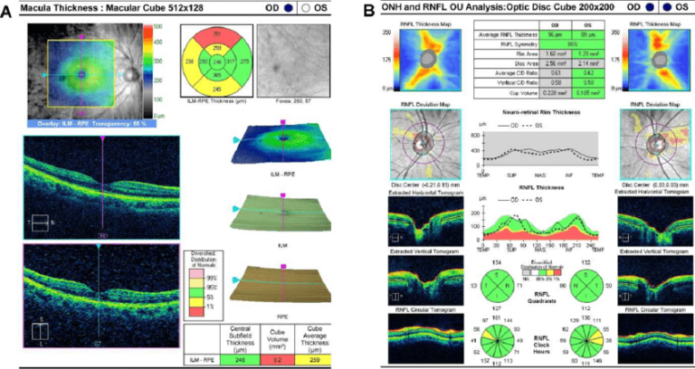
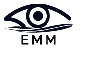
Optical Coherence Tomography (OCT)
Use: OCT is a non-invasive imaging technique that provides high-resolution cross-sectional images of the retina. It aids in diagnosing and monitoring conditions such as macular degeneration and diabetic retinopathy.
Principles: OCT employs low-coherence interferometry to capture detailed images of the eye’s layers. It allows for precise visualization of retinal structures and abnormalities.
Indirect Ophthalmoscope
Uses: The Indirect Ophthalmoscope is a vital tool for examining the peripheral retina and vitreous humor. It aids in the detection and monitoring of conditions such as retinal tears, detachments, and diabetic retinopathy.
Principles: Indirect Ophthalmoscopy involves the use of a condensing lens and a light source to create a wide, stereoscopic view of the retina. The examiner employs a head-mounted light to navigate the beam, enabling a comprehensive examination of the eye’s posterior segment.
Keratometer
Uses: Keratometers are essential for assessing corneal curvature, crucial information for contact lens fitting and refractive surgery planning. They aid in diagnosing astigmatism and monitoring corneal irregularities.
Principles: Based on the principle of reflection, Keratometers measure the size and shape of the corneal image reflected from the patient’s eye. By assessing the cornea’s curvature, these instruments provide valuable data for determining appropriate vision correction.
Fundus Fluorescein Angiography (FFA)
Uses: FFA is a diagnostic tool that uses a fluorescent dye to visualize blood flow in the retina. It is instrumental in detecting and monitoring conditions such as macular degeneration, diabetic retinopathy, and vascular abnormalities.
Principles: FFA involves injecting a fluorescent dye into the patient’s bloodstream. As the dye circulates through the retinal vessels, a specialized camera captures sequential images, revealing details of blood flow and highlighting abnormalities in the retinal vasculature.
Founder of EyesMatterMost- an optometry student who loves talking about eyes. I tend to cover topics related to optometry, ophthalmology, eye health, eyecare, eye cosmetics and everything in between. This website is a medium to educate my readers everything related to eyes.