
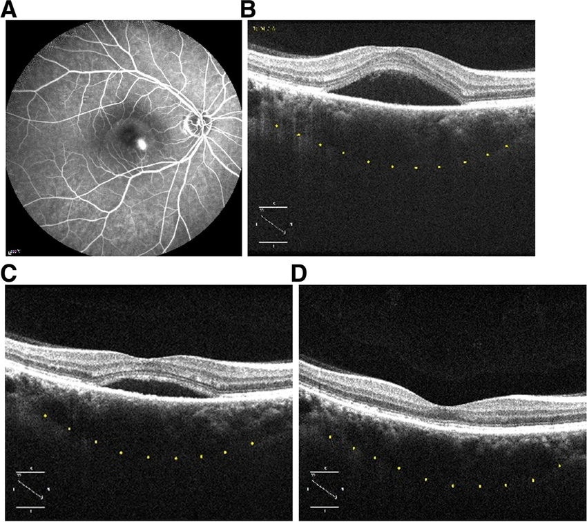
Fluorescein fundus angiography (FFA) uses
Fluorescein Fundus Angiography (FFA) stands out as a diagnostic marvel, offering invaluable insights into the vascular landscape of the eye. This blog explores the diverse and critical uses of FFA, shedding light on how this diagnostic technique aids in the identification, management, and understanding of various eye conditions.
Diagnosis of Retinal Disorders:
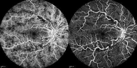
FFA is a cornerstone in diagnosing and characterizing retinal disorders such as diabetic retinopathy. By visualizing the retinal vasculature, clinicians can identify abnormalities, microaneurysms, and areas of ischemia.
Assessment of Age-Related Macular Degeneration (AMD):
In AMD, a leading cause of vision loss in the elderly, FFA helps evaluate the presence of choroidal neovascularization (CNV). It provides crucial information guiding treatment decisions, including anti-VEGF therapy.
Detection of Vascular Occlusions:
FFA is instrumental in identifying retinal vascular occlusions, helping clinicians understand the extent and severity of the blockage. This information aids in formulating appropriate management strategies.
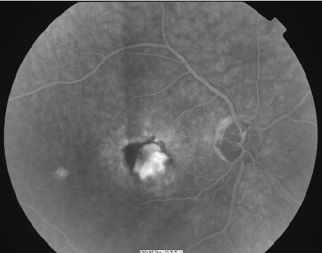
Evaluation of Macular Disorders:
Macular disorders, such as macular edema, are meticulously assessed through FFA. The technique provides high-resolution images, allowing for the identification of fluid leakage and planning targeted interventions. In case of leakage of fluid or blood, there is presence of hyperfluorescence phenomenon.
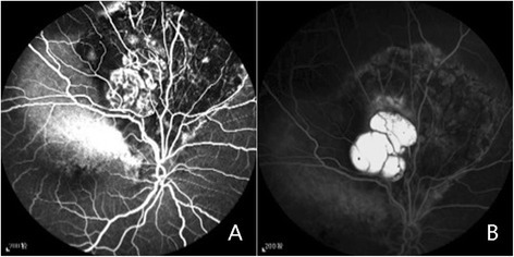
Localization of Tumors:
FFA assists in the localization and characterization of ocular tumors, including choroidal melanomas and hemangiomas. It aids in understanding the vascular patterns within these tumors, contributing to precise diagnosis and treatment planning.
Monitoring Treatment Response:
For patients undergoing treatment for various retinal conditions, FFA serves as a dynamic tool to monitor the response to therapies like anti-VEGF injections or laser treatments. This real-time feedback guides clinicians in optimizing patient care.
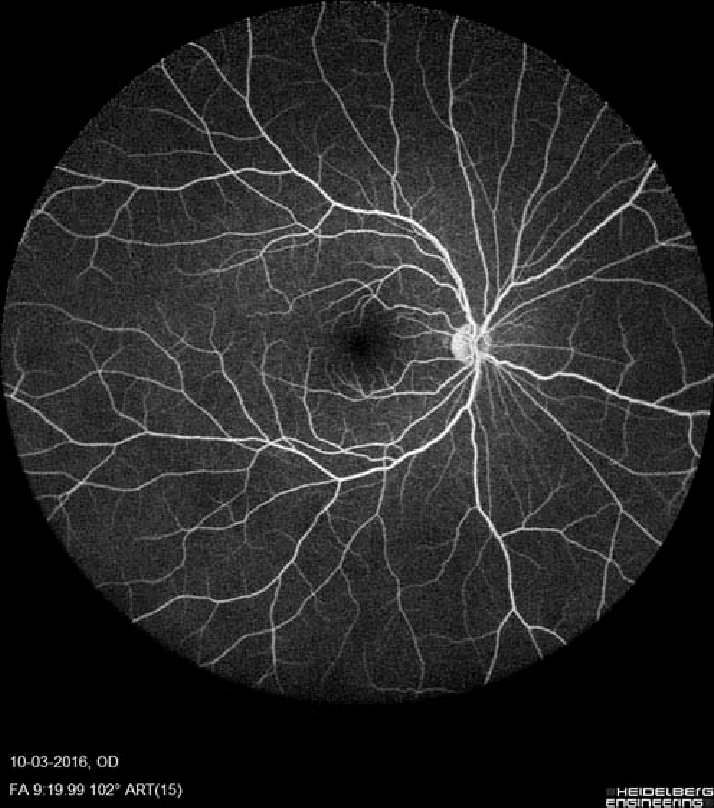
Identification of Optic Disc Disorders:
FFA is used to evaluate blood flow in the optic disc, aiding in the diagnosis of optic disc disorders such as optic neuritis or non-arteritic anterior ischemic optic neuropathy (NAION).
Research and Clinical Trials:
FFA plays a pivotal role in ophthalmic research and clinical trials. Researchers leverage its capabilities to study disease progression, evaluate treatment efficacy, and contribute to advancements in eye care.
Guidance for Surgical Planning:
In certain cases, FFA guides retinal surgeons by providing detailed maps of the retinal vasculature. This aids in surgical planning, ensuring precision during complex procedures.
Early Detection of Abnormalities:
FFA’s ability to detect subtle vascular changes enables early identification of abnormalities, facilitating timely intervention and preventing the progression of conditions that could lead to irreversible vision loss.
Fluorescein Fundus Angiography emerges as a linchpin in the diagnostic arsenal of ophthalmologists. From unraveling the mysteries of retinal disorders to aiding in surgical precision, FFA’s uses are both diverse and pivotal. As technology continues to advance, the role of FFA in safeguarding and restoring vision remains at the forefront of modern ophthalmic practice.
Founder of EyesMatterMost- an optometry student who loves talking about eyes. I tend to cover topics related to optometry, ophthalmology, eye health, eyecare, eye cosmetics and everything in between. This website is a medium to educate my readers everything related to eyes.