
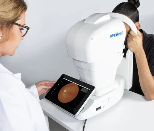
Fundus camera- types, uses and limitations
Fundus cameras play a pivotal role in capturing the details of the eye’s interior. These specialized devices offer a non-invasive means of examining the retina, enabling early detection and management of various ocular conditions.
Fundus camera types
Mydriatic fundus camera
These cameras require dilation of patient’s pupil. These cameras often produce high-resolution images, enabling detailed examination of retinal structures. Mydriatic cameras are commonly used in hospitals and clinics for comprehensive retinal assessments.
Non-mydriatics fundus camera
Non-mydriatic fundus cameras eliminate the need for pupil dilation, making them more patient-friendly. These cameras are particularly useful in settings where pupil dilation might not be feasible or in pediatric cases where patient cooperation is challenging
Hybrid fundus camera
They offer the option to perform examinations with or without pupil dilation, providing flexibility based on the patient’s needs and the level of detail required.
Ultra-Widefield Fundus Cameras
They enable visualization of the peripheral retina, which is crucial for diagnosing conditions like retinal detachments and peripheral retinal pathology. These cameras enhance the clinician’s ability to detect and manage a wide range of retinal disorders.
Uses of fundus camera
The fundus camera is useful to see the fundus and observe changes in it. Here are some conditions and disorders elaborating the importance and use of fundus camera:
- Fundus cameras play a vital role in screening for diabetic retinopathy by capturing detailed images of the retina. These images help ophthalmologists assess the presence and severity of retinopathy.
- Fundus cameras allow for close monitoring of macular changes over time. The high-resolution images produced by these cameras aid in tracking disease progression (Age-related macular degeneration (AMD) affects the central part of the retina, leading to vision loss).
- Glaucoma is characterized by damage to the optic nerve and is a leading cause of blindness. Fundus cameras assist in diagnosing and managing glaucoma by capturing images of the optic nerve head and retinal nerve fiber layer. Specialized imaging techniques, such as optical coherence tomography (OCT) technology integrated into some fundus cameras, provide high-resolution cross-sectional images of the RNFL, facilitating precise measurement and analysis.
- Hypertension can lead to changes in the blood vessels of the retina, a condition known as hypertensive retinopathy. Fundus cameras enable ophthalmologists to visualize these vascular changes.
- Fundus cameras, especially those equipped with ultra-widefield capabilities, aid in identifying retinal detachments by providing a comprehensive view of the retina. This enables swift diagnosis and timely surgical intervention, often preventing permanent vision loss.
- Fundus camera images serve as powerful educational tools for patients. Visual evidence of retinal conditions fosters a deeper understanding of their ocular health. These images also facilitate clear communication between healthcare providers and patients, enhancing treatment compliance and patient engagement with good communication skills.
Top companies manufacturing fundus cameras
Topcon Medical Systems
Topcon is a well-established name in the field of ophthalmic equipment, including fundus cameras. They offer a range of mydriatic, non-mydriatic, and hybrid fundus cameras with advanced imaging technologies.
Carl Zeiss Meditec
They offer fundus cameras with exceptional image quality and innovative features for various clinical applications. Zeiss is a leader in medical technology, known for its cutting-edge optical solutions.
NIDEK
NIDEK specializes in ophthalmic diagnostic and laser equipment. Their fundus cameras are recognized for their precision and versatility in capturing detailed retinal images.
Optos
Optos is renowned for its ultra-widefield imaging technology. They have pioneered the development of ultra-widefield fundus cameras.
Canon Medical Systems
Canon is a global leader in imaging technology, and their fundus cameras offer high-quality retinal imaging solutions for different diagnostic needs.
Other well- known companies for the production of diagnostic equipment in the field of ophthalmology include CentreVue, Kowa Optimed, Clarity Medical Systems.
Limitations of fundus camera
Small pupil in patients
The quality of fundus images is directly impacted by the size of the pupil. Small pupils may restrict the amount of light entering the eye, resulting in limited visibility of retinal structures. Patients with certain medical conditions or discomfort with dilation may pose challenges in obtaining high-quality fundus images.
Medial opacity
Opacity of the eye’s media, such as cataracts or corneal scarring, can significantly hinder the fundus camera’s ability to capture clear images of the retina. These opacities scatter light and compromise the clarity of the retinal image.
Uncooperating patients
Successful fundus imaging requires patients to fixate their gaze steadily and remain motionless during the procedure. This can be particularly challenging, especially when dealing with pediatric patients, individuals with cognitive impairments, or those with conditions affecting their ability to cooperate.
2-D image
Fundus cameras primarily provide two-dimensional images of a three-dimensional structure. Clinicians need to rely on their experience and knowledge to interpret the images and understand the retinal topography.
Peripheral retinal pathology
Peripheral retinal pathology and conditions such as retinal detachments may be missed without a comprehensive view of the entire retina. Ultra-widefield fundus cameras attempt to address this limitation by offering a wider view.
Artifacts
Fundus images can sometimes be affected by artifacts, including reflections, shadows, and distortions.
Cost
Fundus cameras, especially those equipped with advanced technologies, can be costly to procure and maintain. This cost factor may limit the accessibility of fundus imaging, particularly in resource-limited settings or smaller healthcare facilities.
Founder of EyesMatterMost- an optometry student who loves talking about eyes. I tend to cover topics related to optometry, ophthalmology, eye health, eyecare, eye cosmetics and everything in between. This website is a medium to educate my readers everything related to eyes.