
What are drusen of macula?
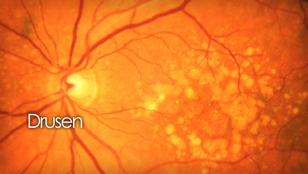
Drusen are the yellowish or whitish deposits which accumulate in different parts of the body. In eyes these deposits are present underneath the surface of the retina. Their presence indicates that the retinal pigment epithelium cells (RPE) cells are not functioning properly. This is the early signs of macular degeneration. The drusen of macula results in distortion and gradual blurring of vision.
The age related macular degeneration is a progressive eye disease that primarily affects older people. It is a leading cause of vision loss in people over the age of 50 specifically in developed countries. AMD affects the macula, which is the central part of the retina responsible for sharp, detailed and central vision. As the disease progresses the size of drusen become larger and leading to thinning and degeneration of the macular tissue. This can result in gradual loss of central vision over time.
Causes for AMD
- Causes of age related macular degeneration which result in drusen are
- Age: individuals over the age of 50, and the risk increases with age
- Certain genetic variations are linked with an increased risk of AMD
- Smoking tobacco products
- Having a close family member with AMD
- AMD is more common in individuals of European descent
Importance of OCT for drusen of macula
The patients of AMD are advised by their ocular physicians to do a test called Optical Coherence Topography (OCT). this is a test to scan retinas. It is a non-invasive imaging technique in ophthalmology to obtain high-resolution cross-sectional images of the retina, optic nerve, and other structures in the eye. OCT provides detailed, real-time visualization of the layers of retina and structures within the eye, allowing for the presence and size of drusen along with integrity of the retinal layers, fluid accumulation and other findings within the macular region.
Types of drusen
Hard Drusen
These are small in size. They appear as tiny yellowish-white deposits beneath the retina. Hard drusen are typically uniform in size and shape. They are associated with normal aging processes. They are often found in older individuals with mild or no vision loss.
Soft Drusen
Soft drusen are larger and more confluent than hard drusen. They are associated with an increased risk of age-related macular degeneration (AMD). They can vary in size ranging from a few micrometers to several hundred micrometers in diameter. They accumulate beneath the retina and specifically in the macula which is responsible for central vision. Early detection and management of soft drusen can help in preserving vision and preventing further vision loss in individuals at risk for AMD.
Cuticular Drusen
Cuticular drusen, also known as basal laminar drusen, present between the retinal pigment epithelium (RPE) layer and Bruch’s membrane. They have a yellowish appearance and may have a “starry sky” or scalloped pattern. Cuticular drusen are associated with a higher risk of developing AMD.
Composition of drusen materials
The exact composition of drusen can vary, but it generally consists of the following components:
- Lipids-they often contain lipids including cholesterol, phospholipids, and fatty acids. Lipids contribute to the characteristic appearance of drusen
- Proteins-complement proteins such as complement factor H (CFH)
- Cellular Waste and Debris- it can contain cellular waste products such as lipofuscin, which is a byproduct of normal cellular metabolism in the RPE
- Minerals: In some cases, drusen may contain mineral deposits, such as calcium deposits resulting in calcification of drusen
Treatment
Modification in lifestyle
Treatment options for diseases associated with drusen appearance may include lifestyle modifications, nutritional supplements, and in some cases, specific therapies to address advanced disease stages. It’s important to note that not all drusen require treatment, and in some cases, the focus may be on monitoring and managing the underlying condition rather than directly addressing the drusen themselves.
Blue-blocking sunglasses
There is no approved treatment to reverse the drusen. The particular physicians recommend the patients to use blue-blocking sunglasses. These are typically brown and polarized sunglasses which block the blue wavelength of light as there is evidence to suggest that excessive exposure to blue light may contribute to the progression of the condition.
These sunglasses help in reducing the amount of blue light reaching the retina, potentially offering some protection to the macula. By filtering out a portion of the blue light spectrum, these sunglasses can help minimize the potential risks associated with prolonged blue light exposure. It is important to choose those glasses which provide sufficient blue light filtration.
Regular examination
Regular eye examinations and monitoring of drusen progression are important to assess changes in the retina and to detect any potential complications or vision-threatening conditions.
Founder of EyesMatterMost- an optometry student who loves talking about eyes. I tend to cover topics related to optometry, ophthalmology, eye health, eyecare, eye cosmetics and everything in between. This website is a medium to educate my readers everything related to eyes.