
Portable slit lamp- things you must know about it
Slit lamp biomicroscope is an instrument designed to observe the eye from cornea to the anterior part of the vitreous and so is the portable slit lamp. They provide magnified view of the parts of the eye illuminated by intense beam of light. One can adjust the intensity and angle of the beam of light. The portable slit lamp is a device which is portable and easy to take it to the patients.
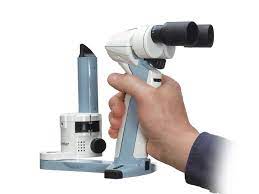
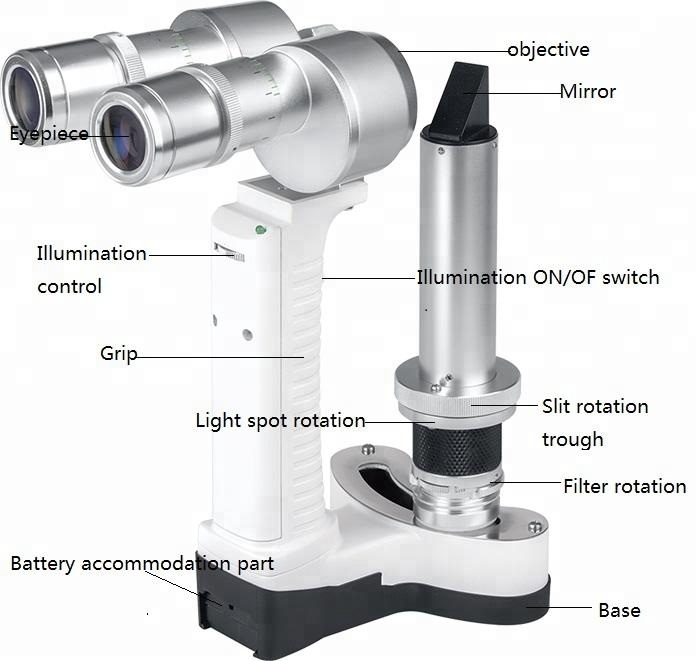
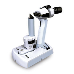
Parts of portable slit lamp
This device comes in aluminum case. The portable slit lamp devices are made up of following parts:
Battery
These are mostly rechargeable lithium-ion batteries. They mostly offer high-energy density, longer battery life, and the ability to be recharged multiple times. The fully charged slit lamp can be used up to 6 hours straight. Other type of batteries used are alkaline batteries, NiMH batteries and nickel-cadmium batteries.
Forehead supporter
The forehead supporter is helpful in maintaining proper alignment and positioning of the patient’s eye.
Focusing plates and microscope
The focusing plates in portable slit lamps help in adjusting width, height and beam angle of the slit lamp beam. Slit lamps generally have a fine focus control mechanism, typically located on the microscope or the slit lamp arm. The microscope on the slit lamp usually has a separate focus adjustment mechanism. This allows the examiner to fine-tune the focus of the microscope specifically for their own visual acuity to ensure clear view of the eye.
Charging docking station
The charging docking station serves as a convenient and dedicated platform for recharging the batteries of the portable slit lamps making it very portable.
Filters
They have interchangeable different type of filters to modify the color or wavelength of the light. Common filters include cobalt blue, red-free, and yellow filters, which enhances specific features to detect certain eye conditions.
Eye pieces
Eye pieces provide the examiner with a magnified view of the patient’s eye with depth perception for accurate assessment and examination of the eye. They often have dioptric power adjustment settings that enable the examiner to focus clearly while examining the eye.
Uses of portable slit lamps
The portable slit lamp examination includes the observation and diagnosis of following conditions and underlying disorders:
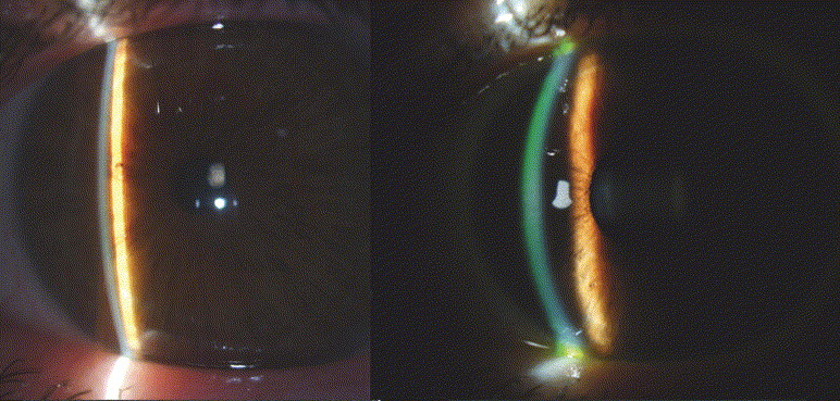
Thickness of cornea
Corneal thickness is an important factor in determining risk of glaucoma, corneal edema due to accumulation of fluid in different corneal dystrophies and diseases, corneal refractive surgeries as thinner cornea may have limitations in getting LASIK or PRK, corneal disorders, infections, corneal trauma and contact lens fitting.
Anterior chamber depth
The anterior chamber depth helps in observing and assessing the angle between the cornea and the iris known as the anterior chamber angle. The shallow depth of anterior chamber indicates disposition to the risk of angle-closure glaucoma.
If the anterior chamber depth is abnormally shallow, it may suggest that the lens is displaced forward. This can occur in conditions such as lens dislocation or certain types of cataracts. The anterior displacement of lens occurs in conditions like Marfan Syndrome, trauma, ectopia lentis, connective tissue disorders like Ehlers-Danlos syndrome. This can happen if there is damage to the zonules during the surgical procedure.
Size of pupil
The size of pupil is controlled by the autonomic nervous system. The abnormal size of pupil (persistent dilation or contraction or any asymmetry may indicate underlying medical conditions or neurological abnormalities like Anisocoria, Horner’s Syndrome, Adie’s tonic pupil, oculomotor nerve damage resulting in dilated pupil (mydriasis) and limited eye movements.
Endothelial cell count
The endothelial cell count gradually decreases with age due to decrease in cell replication capacity. Monitoring endothelial cell count in older patients helps to assess the extent of age-related changes and distinguish them from pathological conditions.
Presence and type of cataract
The slit lamp examination allows the ocular physicians to examine the cataract’s presence and location within the lens. In nuclear cataract, the opacity and lenticular changes appear in the center of the lens. Cortical cataracts appear as white, wedge-shaped opacities extending from the periphery of the lens towards the center. Posterior subcapsular cataracts occur at the back surface of the lens, just beneath the capsule. In this type of cataract, the slit lamp examination may show as irregularly shaped opacities at the back of the lens.
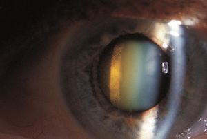
Posterior subcapsular cataract formation shown in slit lamp examination
Intra-ocular pressure
Slit lamp exams are used to measure intraocular pressure (IOP) of the eye through a procedure called tonometry.
Contact lens fitting
The contact lens fitting is very important for the permeability of air and oxygen for the surface of the eye. Any maladjustment can result in hypoxia which can also lead to development of corneal ulcers in worst case scenario. The slit lamps allow ocular physicians to do initial lens assessment, adjustment and evaluation and corneal health assessment.
Companies to purchase portable slit lamps
Here are a few well-known companies that specialize in ophthalmic equipment and are often considered reliable options for purchasing portable slit lamps:
Topcon is a globally recognized company in the field of ophthalmic equipment. They offer a range of portable slit lamps known for their best quality.
Haag-Streit is a renowned manufacturer of ophthalmic instruments including slit lamps. They have a reputation for producing high-quality slit lamps that are widely used in clinical practices.
Keeler is a reputable name in the ophthalmic industry which offers a variety of portable slit lamps.
Reichert is a well-established company that produces a range of ophthalmic instruments.
Founder of EyesMatterMost- an optometry student who loves talking about eyes. I tend to cover topics related to optometry, ophthalmology, eye health, eyecare, eye cosmetics and everything in between. This website is a medium to educate my readers everything related to eyes.