
Contrast sensitivity- all crucial aspects
The ability of the eye to determine the crispness of the image against its background is known as contrast sensitivity. It is a measure of how well an individual can perceive differences in luminance or color of the adjacent areas of an image.
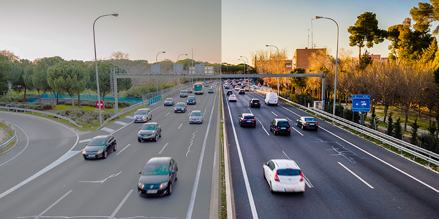
It is an important aspect of visual functions because it is also considered along with visual acuity while evaluating an individual’s visual performances. Thus it helps in determining the appropriate corrective measures or interventions, such as glasses, contact lenses, or surgical procedures. The human visual system is more sensitive to contrasts in certain spatial frequencies than others.
Types
Spatial Contrast Sensitivity:
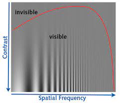
Spatial CS refers to the ability to perceive contrasts in different spatial frequencies. The humans tend to have higher sensitivity to medium spatial frequencies, which correspond to moderate-sized details or patterns in the visual field.
Temporal Contrast Sensitivity:
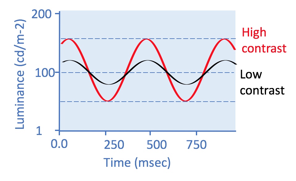
Temporal CS relates to the sensitivity of contrast in dynamic or time-varying visual stimuli. It refers to the ability to sense changes in contrast over time. Temporal contrast sensitivity is important for perceiving moving objects, tracking motion, and distinguishing objects from their background in motion.
Chromatic Contrast Sensitivity:
Chromatic CS involves the perception of contrast differences in color. The visual system is capable of distinguishing variations in contrast between different colors or color combinations and it also involves the extent of saturation of certain color.
High-Contrast Sensitivity:
This involves distinguishing objects from their background when the contrast between the object and the background is prominent.
Low-Contrast Sensitivity:
Low-CS involves the ability to perceive small differences in contrast between object and its background.
Parts of brain involved in CS
These are the areas of brain involved in generating contrast sensitivity:
Primary Visual Cortex (V1), secondary visual cortex (V2), the dorsal stream (including the posterior parietal cortex), the ventral stream (including the inferior temporal cortex), Lateral Geniculate Nucleus (LGN), Superior Colliculus, Frontal and Parietal Lobes
The human brain is a vast in its functions and structures. The involvement of specific brain regions involved in generating contrast may vary depending on the specific task, context, and visual stimulus being processed.
How to measure contrast sensitivity
Here are a few commonly used methods or charts to measure CS:
Pelli-Robson Contrast Chart:
This is a widely used chart that consists of several rows of letters, each row having decreasing contrast. The individual is asked to identify the letters on the chart up to the lowest contrast level he can read. Its working distance is 1m.
Freiburg Visual Acuity & Contrast Test (FrACT):
This is a computer-based test that assesses both visual acuity and contrast sensitivity of the patient.
CSV-1000E Contrast Sensitivity Test:
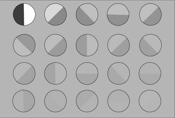
The CSV-1000E is a device that uses sine-wave gratings to assess contrast sensitivity. The individual views the gratings at different spatial frequencies and contrasts.
Melbourne Edge Test:
This test helps in measuring contrast sensitivity of low vision patients.
Mars Letter Test:
This test employs specially designed letters that have varying contrast levels. This chart is almost similar to Pelli- Robson chart but smaller in size.
CS measurement
Some common ways to measure contrast sensitivity are given below:
- It is measured in optotypes with varying contrast levels to the individual. The letters are typically arranged in rows, and the person being tested identifies or reads the letters.
- It is also measured in sinusoidal gratings with varying spatial frequencies and contrast levels. The patient is asked to identify the orientation of the grating (e.g., vertical or horizontal) or detect the presence of the grating.
- We can measure it using specialized computer software or digital device as they have typically present stimuli such as gratings, letters, or images with varying contrast levels.
- Functional Vision Analyzer- a device that measures various aspects of visual function, including contrast sensitivity. It presents gratings or other visual stimuli with different spatial frequencies and contrasts.
Factors affecting contrast sensitivity
These are some non-ocular or systemic causes that decrease CS
- Age, it is said that CS decreases 10 percent with every decade of life.
- Diabetes mellitus
- Hypertension
- Multiple Sclerosis
- Pituitary adenoma
Ocular factors affecting CS
- Corneal haze
- Lenticular changes
- Cataract
- Glaucoma
- Refractive surgeries
- Ocular hypertension
- Retinitis pigmentosa
- Amblyopia
- Keratoconus
- Retrobulbar neuritis
Founder of EyesMatterMost- an optometry student who loves talking about eyes. I tend to cover topics related to optometry, ophthalmology, eye health, eyecare, eye cosmetics and everything in between. This website is a medium to educate my readers everything related to eyes.