The science of vision- How do our eyes work?
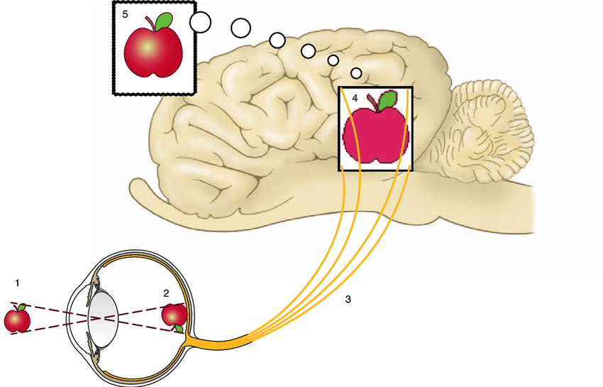
The science of vision- How do our eyes work? Let’s make it as simple yet comprehensive as it can be for every little biologist or anybody who gets fascinated by the fact that these tiny yet paramount organs of vision are working and making us see the nature, our friends, the great architects, observe the patterns of fingers while playing musical instruments and so on. What is the science of vision. And how do our eyes work and let us see the micro-organisms under microscope and celestial bodies clearly through telescopes.

Refraction- bending of light
Lens
Our eyes are the refractive organs and our each eye has power of almost 58 to 60 diopters. Our lens and cornea have these powers and when the light pass through both of them, it refracts and falls on the retina.
Cornea
First the light passes through the cornea which has a power of almost +43 diopters and it bends and passes through the pupil. The size of pupil depends upon the intensity of light and extent of work you are performing e.g. pupil dilates in near work.
For near work
After refraction through cornea the light rays converge as now they have to pass through another refracting part of the vision organ- the eye. Now the light rays will pass through the lens of the eye. The lens has a power of +16 to 20 diopters while doing distant work. However if you perform near work, then the shape of the lens changes which changes its power which becomes now 33 diopters.
Wider central vision
The light passing through the center of the nucleus of lens gets straight to the retina. All the light rays coming through the periphery do not get through the refraction process and hence we get clearer central view a limited range of peripheral view.
The retina- curtain of the eye
The retina of the eye is known as the curtain of the eye. The special cells in retinal help us see the color vision, contrast and depth perception. The rods and cones cells in the retina of eye have special pigments which undergo biochemical changes when light falls on them.
Optic nerve and chiasma
These changes stimulate the neural layers of the retina to stimulate the visual pathway to send the message up to visual cortex. The axons of ganglion cells of the retina form the optic nerve. The optic nerve carries these signals up to optic chiasma.
Optic tract
The optic chiasma is the structure where the fibers coming from the nasal half of the retina through optic nerve decussate and pass to the optic tract of opposite sides. The fibers from the temporal half of the retina coming through the optic nerve however to the optic tract of the same side.
Lateral geniculate body
The nerve fibers from the optic tract pass to the lateral geniculate body. The fibers through the lateral geniculate body travel to the visual cortex (occipital cortex) through optic radiations.
Visual cortex- master of vision
The visual cortex of the brain receives the information coming through the retina of the eye and processes it to make us see erect image. The image formed on the retina is real, diminished and inverted. It is the visual cortex of the brain that processes this information and makes us see the straight image of exact same size.
Visual cortex importance
The visual cortex of the brain is further divided into different layers and perform the critical functions related us to see colors, depth, contrast and motion of the bodies. Any damage to the certain part of the visual cortex an effect its associated function.
Founder of EyesMatterMost- an optometry student who loves talking about eyes. I tend to cover topics related to optometry, ophthalmology, eye health, eyecare, eye cosmetics and everything in between. This website is a medium to educate my readers everything related to eyes.

