Ultrastructure of extraocular muscles
Let’s give a quick overview before understanding the ultrastructure of extraocular muscles. These muscles of the eye help us attain binocular single vision and stereopsis of the image. Both the eyes have them and they work in synchronization with each other. They maintain the visual targets. It is an art to use both eyes at the same time to see the world. The normal functioning of these muscles play role in it. They are responsible for precise and rapid movements of eye.
Extra ocular muscles are the special type of skeletal muscles (the muscles whose movements can we control on our own). In extra-ocular muscles (EOM), not all the fiber types propagate action potential unlike skeletal muscles.
Names
Following are the extraocular muscles:
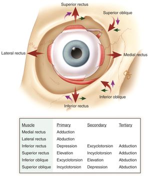
- Medial rectus
- Lateral rectus
- Superior rectus
- Inferior rectus
- Superior oblique
- Inferior oblique
- Levator palpebrae superioris (this muscle gives extra 2mm elevation to the eyelid in angry state)
Here we will only talk about recti and oblique muscles. Each EOM is composed of two layers: orbital layer and global layer.
Orbital layer
The orbital layer is closer to the orbit of the eye and ends before the muscle becomes tendinous at the global layer.
Global layer
The global layer is adjacent to the eyeball and it inserts into the eye through a tendon.
Ultrastructure of extraocular muscles
Before digging the ultrastructure, it is important to understand that a muscle is made up of muscle fibers. Muscle fibers have myofibrils that run parallel in the muscle fiber. The myofibrils are made up of contractile protein actin and myosin proteins. The ultimate contractile unit of skeletal muscle and extraocular muscle lies in myofibrils.
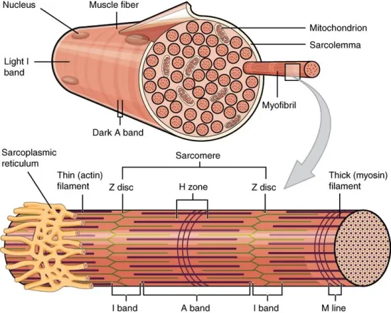
There are thick and thin filaments that give the muscle fiber a striated appearance. The thin filaments are composed of actin proteins and thick filaments in the myofibril have myosin proteins. The thick filaments make A-band (dark band) and thin filaments make I-band (thin band). The imaginary line at the I-band is called Z-line. The A-band is surrounded by two I-bands as shown in the figure.
Sarcomere
The difference between adjacent Z-lines constitutes the contractile unit of the muscle called sarcomere. The contraction of sarcomere results in the contraction of myofibrils which in turn contracts muscle fiber and eventually the muscle. It is the functional unit of muscle.
Breaking and formation of cross-bridge
The breaking and formation of cross bridges result in the hydrolysis and formation of ATP which drives the sliding of actin protein towards the myosin head. This results in shortening and contraction of sarcomere.
How nerve innervation derives contraction of muscle?
The cell membrane of skeletal muscles is known as sarcolemma. There is an invagination of sarcomere at the A-I junction at the ultrastructure level. The tubule like structure is in contact with extra cellular matrix. These membranes have Calcium channels which open when the membrane receives action potential. The cisternae of sarcoplasmic reticulum adjacent to the T-tubule on both sides make a configuration called triad. The cisternae are filled with a dense granular material containing calsequestrin, a protein having a high affinity for calcium.
The opening of calcium channels result in the entry of Ca2+ ions in the cell and eventually they attach with a protein called topomyosin. The attachment of topomyosin with Ca2+ ion results in the exposing of actin binding site on myosin. Thus the site of cross bridge formation is exposed and all ready to make cross bridge formation.
More contractile units
Extraocular muscles have more contractile units than skeletal muscles and their ratio of nerve innervation to muscle fiber is in the range of 1:3 to 1:5 which is greater than that of skeletal muscles, 1:50 to 1:125.
Extraocular muscle fibers classification
The extraocular muscle fibers are classified as following
- Orbital single innervated fiber (most fatigue resistant)
- Orbital multiple innervated fiber
- Global red single innervated fiber (highly fatigue resistant)
- Global intermediate single innervated fiber (intermediate level of fatigue resistance)
- Global pale single innervated fiber (low fatigue resistant)
- Global multiple innervated fiber
These fibers are classified on the basis of type of muscle fibers, single or multiple nerve innervation and mitochondrial pattern. The red muscles have highest content of mitochondria than intermediate and pale fibers. The highly fatigue resistant fibers are fast twitch fibers and slow twitch fibers are less fatigue resistant.
Founder of EyesMatterMost- an optometry student who loves talking about eyes. I tend to cover topics related to optometry, ophthalmology, eye health, eyecare, eye cosmetics and everything in between. This website is a medium to educate my readers everything related to eyes.