
Why we get with and against reflex movements in retinoscopy
Why we get with and against reflex movements in retinoscopy? Wither of this scenario depicts the ametropic conditions of the eye which are needed to be given right prescription for the refraction.

What is a Retinoscope?
Retinoscope is an instrument which is useful for the objective refraction of the patient. This instrument helps in determining the refractive error of the patients.
Concept of working distance
We focus the beam of light from the retinoscope into the patient’s eye. The ideal way is to sit at the far point of emmetrope and then focus the beam of light into the patient’s eye. This is inconvenient for the examiner to get from his seat and change lenses in the trial frame of patient until the error neutralizes. We sit at a distance of our arm length in front of the patient and while doing this we have practically shifted the far point of the patient in front of his eyes.
This reciprocal of our arm length (if the arm length of the examiner is 67 cm then its reciprocal is 1/67= 1.5D) is the amount of refractive error that has to be subtracted from the neutralization values. The arm length varies person to person but the ideally accepted average length is 67cm.
Streaks projecting from retinoscope
The most commonly used streaks or beams of light from retinoscope are vertical and horizontal streaks. Consider a retinoscope in your hand and you choose vertical streak. Now remember that your hand movement with this streak is in horizontal direction. Similarly for the horizontal streak the hand movement is in vertical direction.
Calculations
While performing calculations and drawing meridians for calculations, consider the direction of our hand movement while preforming retinoscopy i.e. the horizontal hand movement with vertical streak corresponds to noting the measurement of neutralization value of lens in the horizontal meridian direction and vice versa. The whole game depends upon the neutralization of the error. Neutralization is also called end point of retinoscopy.
Pupillary reflex
While observing the patient’s eye with the help of retinoscope, we get red reflex (image of retina in the patient’s own pupil corresponding to normal retina.
Examining with and against reflex movements in retinoscope
Now after adjusting the type of streak (vertical, horizontal or oblique) the examiner sits at an arm’s length in front of patient and observes the behavior of reflex while moving the retinoscope.
If the pupillary reflex does not move at all with the movement of retinoscope then the error is neutralized i.e. there is no movement of pupillary reflex in the eyes.. If the pupillary reflex moves along with the movement of streak of light directed from retinoscope, then there are following possible conditions:
- Myopia lesser than 1.5D
- Hypermetropia shows with movement
- Emmetropia can also show with movement
- Reasons of formation of with reflex
The ray diagrams are very helpful to show the exact position of projection of light (vertical/horizontal streak of light). The command over why we get with and against reflex movements with retinoscope can help in treating the patients refractive error more efficiently.
Reason of reflex movements with ray diagrams
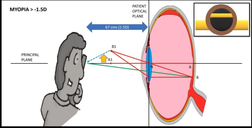
Myopia lesser than 1.5 D
The inverse of power is the focal length. If the examiner is at a distance of 67 cm, then the myopia less than 1.5 D will have far point behind the examiner (at a distance greater than 67 cm). consider the diagram. The central retinal point i.e. A is projected to point A1 along the principle plane. A1 is the far point of the patient. Now the examiner looks downward in the eye of patient in para-central point B on the retina is projected at point B1 at the far point of the patient.
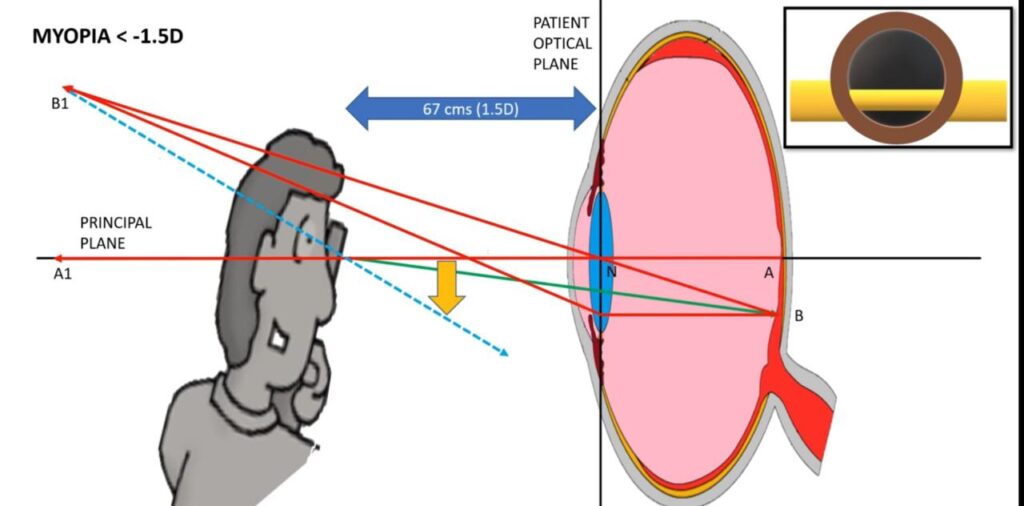
Now when the blue rays extend from the point B1 to the retinoscope, their relative position to the retinoscope is downwards. This shows pupillary reflex in WITH- MOVEMENT direction. This gives movement in the same direction of retinoscope. The position of blue dotted rays with respect to principle plane indicates the movement of pupillary reflex.
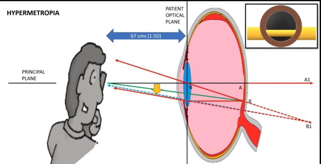
With reflex in hypermetropia
In hyperopic patients, the far point forms virtually behind the retina which is A1. The paracentral point B is projected at B1 through diverging rays. On extending the blue arrow, the relative position of blue arrow to the principle plane is downward. This causes WITH-MOTION pupillary reflex in hyperopia. i.e. the reflex moves along the direction of retinoscope’s streak.
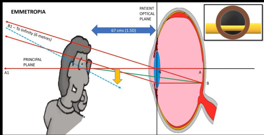
With movement in emmetropes
In emmetropes as per the figure, the far point is at infinity and when the rays extended from point B1 to the retinoscope, they are downward with respect to principle plane when the examiner looks downward in the patient’s eye. This shows with movement.
In myopes of the error of 1.5D, their far point is equal to the working distance of the examiner’s arm length. At this point the examiner cannot correctly predict the with and against movement of the reflex and the reflex seems neutral.
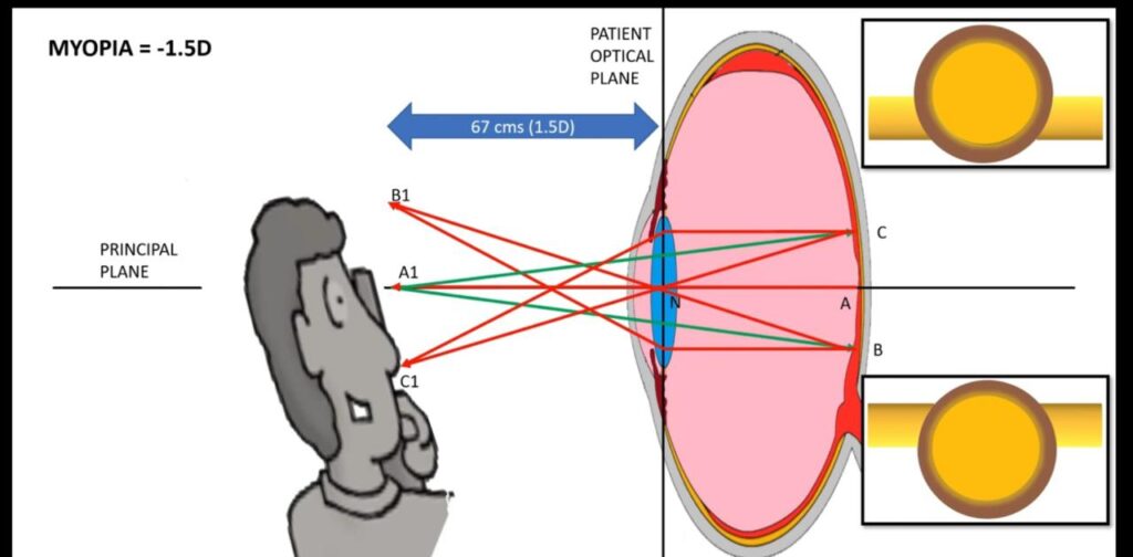
Against movement of pupillary reflex
The far point of these myopes is closer than the working distance and the rays projecting from B converge at B1. When the rays at B1 are projected at the retinoscope, we see against motion with respect to the principle plain. This is the phenomenon of against reflex movement.
Founder of EyesMatterMost- an optometry student who loves talking about eyes. I tend to cover topics related to optometry, ophthalmology, eye health, eyecare, eye cosmetics and everything in between. This website is a medium to educate my readers everything related to eyes.