
Things you must know about uveal tract
The word “uvea” is derived from Latin word “uva” which means grapes. If we remove sclera and observe the eye then its choroid appears to be grape shaped structure.
Coats of the eyeball
The eyeball has three coats:
- The outermost coat is made up of sclera and cornea
- The middle coat is uveal tract
- The innermost coat is the retina of the eyeball .
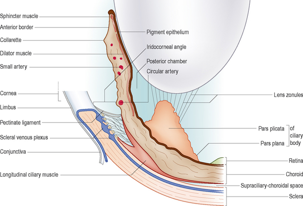
Uveal tract
The uvea or uveal tract is the middle layer of the eyeball. It is highly vascularized and it has three parts which are choroid, ciliary body and iris.
Uvea can be summarized as densely pigmented middle vascular coat of the eyeball. It extends from the anterior aspect of the globe of the eyeball all the way to the posterior aspect of the globe at the optic nerve.
It receives blood from the anterior and posterior ciliary branches of ophthalmic artery. The uveal tract is very pivotal in providing the vasculature and nourishment to the eye.
Choroid- posterior part of uveal tract
It is the posterior part of the uveal tract. The choroid is highly vascularized structure. It has following parts:
Suprachoroidal lamina
the layer of choroid facing sclera is known as suprachoroidal lamina. It has macrophages, fibroblasts and collagen fibers.
Stroma of choroid
It is the loose connective tissue containing pigment cells, macrophages and plasma cells. It is very rich in blood vessels. The presence of blood vessels clearly depicts its function that it provides nutrition and oxygen to the interior of the eye.
Bruch’s membrane
It is the layer of choroid which is in contact with the pigmented epithelium of retina.
The thickness of choroid decreases with aging. It also provides nutrition to the retina up to its plexiform layer. The decrease in the thickness of the choroid can affect its function of providing nutrition and oxygen to the retina. It can cause age related macular degeneration.
Ora seratta
Ora seratta The junction between choroid and ciliary body is known as ora seratta.
Ciliary body
The vascularity of the middle coat of eye continues to grow to the anterior side of the choroid in a thick part which is known as ciliary body. These are triangular in shape. It has two parts. The posterior part of ciliary body that continues with choroid is plane and hence it is pars plana .The anterior part of ciliary body has many folds in it and this is called pars plicata.
Ciliary body has three major parts:
- Ciliary muscles
- Ciliary body stroma
- Ciliary epithelium
Beneath ciliary muscles there is highly vascular stroma. It is lined by non pigmented ciliary epithelium. This structure collectively forms loop like structure which are ciliary processes.
Function
The ciliary process secretes aqueous humor in the posterior chamber of eye (between lens and posterior aspect of iris) that circulates through it and anterior chamber of the eye.
Composition
Water, ions (sodium, chloride and bicarbonate) amino acids, glucose and ascorbic acids form the aqueous humor.
The ciliary body plays role to keep the eye distended to achieve its optical functions. This makes the intra-ocular pressure of the eye. It also provides nutrients and carry waste products from the avascular structures of the eye which are lens and cornea. It is clear so that light can pass through both the lens and cornea to fall on the retina.
Iris- anterior most part of uveal tract
The anterior most part of uveal tract is iris. It is circular diaphragm structure with an aperture of about 4mm. This aperture is pupil. It attaches to the anterior surface of ciliary body. The iris separates cornea and lens making anterior and posterior chamber of the eye.
Iris has two zones:
- Pupillary zone
- Ciliary zone
It has 5 layers:
- Anterior limiting layer
- Stroma (contains melanocytes which determine the color of eye)
- Muscular layer (Smooth muscle at pupillary margin and in iris stroma)
- Anterior pigment epithelium
- Posterior pigment epithelium
Function of iris
The two types of muscles that mainly form the function of iris are sphincter pupillae and dilator pupillae. The sphincter muscle causes constriction of pupil and limits excessive light to enter and damage the eye. It has parasympathetic innervation. The dilator pupillae has sympathetic innervation and it causes dilation of the pupil and let more of the light to enter the eye to make clear and visible image on the retina. It regulates the amount of light entering the eye.
What is Uveitis?
The inflammation of uveal tract is known as uveitis. As uvea is not a single organ or parts it is very profound structure.
Types of uveitis
The anatomical types of uveitis are following
- Anterior uveitis (iris gets affected e.g. iritis, Iridocyclitis)
- Intermediate uveitis (affects the vitreous gel)
- Posterior uveitis (affects choroid and retina of the eye)
- Pan uveitis (inflammation of all parts of uveal tract)
Symptoms
The common symptoms of uveitis include
- Pain
- Photophobia
- Redness
- Blurredness
- Floaters
Decrease in vision The ocular physician confirms the diagnosis after looking the interior of the eyeball by dilating the pupils.
Founder of EyesMatterMost- an optometry student who loves talking about eyes. I tend to cover topics related to optometry, ophthalmology, eye health, eyecare, eye cosmetics and everything in between. This website is a medium to educate my readers everything related to eyes.