Extraocular muscles and movements
It is said that there is more anatomy in per cubic centimeter in the orbit of eyeball than anywhere else in the body. But all we need is better explanation and easy way to grasp the knowledge to apply in the medicine. Lets discuss about the extraocular muscles and the movements they cause
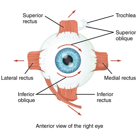
There are six muscles that move the eyeball. Let’s decode the information about the muscles and their arrangement that synchronize the movements of the eyeball as a whole. These extraocular muscles are next to the bony walls of the orbit. There are four straight muscles and two oblique muscles in each eye which make extra-ocular muscles of each eye.
Extraocular muscles
The four muscles are straight and they are:
Superior rectus (SR)– it attaches to the orbit of eye superiorly
Inferior rectus (IR) – it attaches to the orbit of the eye inferiorly
Media rectus (MR)– it attaches to the orbit on medial side (nasal side)
Lateral rectus (LR)– it attaches to the orbit on the lateral part of the eyeball
The two oblique muscles are:
Superior oblique muscle (SO)
Inferior oblique muscle (IO)

The recti muscles are straight muscles and their muscle fibers are running straight to each other.
Superior oblique muscle
The superior oblique muscle of the eye comes from apex of the orbit and attaches to the eyeball on the anterior-medial side and it passes through the trochlea and it runs posterior-laterally and inset into the eyeball whereas trochlea is a pulley like tendinous structure present in the superior nasal aspect of the frontal bone.
All the recti muscles of the eye along with superior oblique muscle come from common tendinous ring
Inferior oblique muscle
It is present on the inferior side running and curving from medial to lateral around the eye. Its origin is maxillary bone.
Movements associated with extraocular muscles
The extra ocular muscles help eyes to move along three different axes
- The horizontal axis– along the horizontal axis the movement of eyeball is up and down
- The vertical axis– this allows movement in left and right direction which gives rise to medial and lateral movements of eye if the right eye is under consideration.
- The anterior-posterior axis i.e. along the mid of the orbit; it allows the eyeball to rotate.
Lets talk about the uniocular movements of the eye
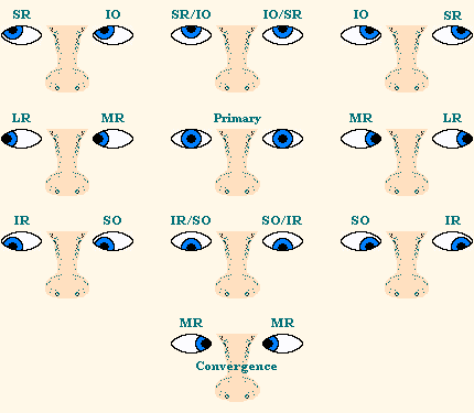
The movements in one eye with the help of extra-ocular muscles are these movements. These are called duction movements. There are six types of these movements
Abduction and adduction
The movement of the pupil of eye towards the lateral side is called abduction whereas the movement of the eyeball towards the medial position is called adduction. It’s quite like the abduction and adduction of the limbs.
Muscles
The lateral rectus (LR) when contracts causes abduction and it is the primary abductor and medial rectus causes adduction hence also known as primary adductor.
Supraduction and infraduction
The movement of eyeball upward i.e. when pupil rises is called supraduction or elevation. On the contrary, the movement of the pupil downward is called infraduction or depression.

Muscles
Superior rectus (SR) causes supraduction or elevation as its primary role. It also causes elevation of the abducted eye. Besides its primary function it also causes limited adduction and intorsion of the eye. It is also responsible for the adducted depression of the eye.
The inferior rectus causes infraduction or depression. It is also responsible for the elevation of adducted eye. They are most effective in causing abducted depression of the eye. It also helps in adduction and extorsion of the eye.
The contraction of any of these recti muscles cause the movement of the eye in their respective direction as these muscles pull the eye.
The insertion of both inferior and superior rectus in the eyeball is such present in a way that if they both contract then they can pull the eyeball medially. Hence they can cause adduction as well.

Intorsion and extorsion
Consider you are observing right eye. If you rotate the right eye medially then it is intorsion.
If you rotate the same eye laterally then it is lateral rotation or extorsion.
Inferior oblique muscle’s primary function is the extorsion of the eye. It also causes elevation of fully adducted eye. It is the only effective elevator in the fully adducted eye. It also contributes in abduction of the eye.
Importance of extorsion and intorsion
The rotational movements i.e. intorsion and extorsion help the eyes to keep the image of the objects stable and on the same position on the retina. So that the binocularity with depth perception can be achieved without the objects moving along with the tilting of our head. When you tilt your head suppose you are looking at the arrow with arrow head pointing superiorly then the image does not move along with your head tilting. You are tilting your head on the left direction. In this case the right eye is intorted and the left eye has been extorted equally. This helps the image to fall on the same part of retina.
Founder of EyesMatterMost- an optometry student who loves talking about eyes. I tend to cover topics related to optometry, ophthalmology, eye health, eyecare, eye cosmetics and everything in between. This website is a medium to educate my readers everything related to eyes.
