Hypermetropia- things you must know about it
Hypermetropia
Hypermetropia or hyperopia is the type of refractive error in which the image of the object is formed behind the retina. It is also known as farsightedness. The people who have hyperopia cannot see distant objects if their accommodation is at rest. However they can see far if their lenses accommodate to make the image fall on retina. The phenomenon of accommodation occurs with the help of ciliary tone.
Generally with the age the ciliary tone decreases. Hence the far objects cannot be seen clearly with the help of accommodation. The ciliary tone of the muscles can accommodate up to 1 diopter. It means that it can cover hyperopia up to 1 diopter naturally. Hyperopia generally affects near vision making the vision blur.

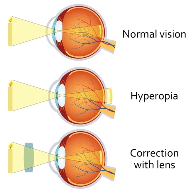
Image formation in hypermetropia
In the eyes as the size of eyeball is short the image is formed behind the retina. At birth, the babies have small eyeball due to which they are also hyperopic. Their hypermetropia decreases with the passage of time when their axial length and eyeball grows in a process called Emmetropization.
If the eyeball parts and axial length do not grow to a normal range then the children can have hyperopic shift in their eyes. If their eyeball grows beyond normal then a child acquire myopic shift in his eyes. By the age of 5 to 7 years the hyperopia generally goes away. After the age of 40 years the chances of farsightedness increase because of age related changes in the lens. The eyes also lose the ability of accommodation due to changes in lens as it becomes thicker. It is called presbyopia. The prescription for presbyopia varies depending upon the age. The people of age 40 are generally prescribed +1 D binocularly. It can be up to +3 Ds in older people.
Presbyopia is an accommodation error and hyperopia is a refraction error. Presbyopia occurs in senile age.
Grading of hypermetropia
According to American Optometric Association the hypermetropia has three grades:
- Low hypermetropia (<+2Ds)
- Moderate hypermetropia (>+2 and <+5Ds)
- High hypermetropia (>+5Ds)
Symptoms
The person can be asymptomatic
- The symptoms include eye strains with or without vision loss, watering, mild photophobia and headache.
- These symptoms occur because the accommodation is not at rest even looking at the far objects.
Causes
- Shortness of axial length of the eye- according to the book Theory and Practice of optics and refraction by AK Khorana the 1 mm shortening of axial length can induce 3 diopters of hypermetropia.
- Flattening of curvature of cornea- this can result in less bending of the light rays that instead of focusing on the retina focus behind it.
- Index hypermetropia- it occurs due to decrease in refractive index of lens due to its sclerosis
- Aphakia – a condition in which the lens is not present in the eye. It can be congenital or acquired
- Positional hypermetropia- the disposition of lens due to congenital or acquired reasons and the acquired reasons can be a trauma to the eye.
Clinical findings
- Small axial length
- Cornea appears smaller
- Shallow anterior chamber– lesser than 2.4mm
- Retinoscopy or auto refraction reveals hyperopic shift of the eyes
- Fundoscopy reveals small optic disc. It is also called pseuodpapillitis because there is no actual swelling of the optic disc.
- Shot silk appearance of retina– it occurs because of the short eyeball the retina is nearer to the examiner due to which it shines brighter with greater brilliance
Classification
It has three main classes
Physiological hypermetropia
It is also known as simple hypermetropia. It occurs when the axial length of th eyeball grow behind normal biological growth.
Non physiological or pathological hypermetropia
- Congenital
- Acquired
Congenital causes include micro-cornea, micro-ophthalmous, aphakia and posterior sub-luxation of lens.
Acquired hyperopia occurs due to aphakia, posterior sub-luxation, senile and positional hypermetropia. These symptoms develop later in life e.g. sub-luxation and aphakia conditions occur due to trauma.
It can also occur due to over correction of myopia. The doctors overcorrect myopia by flattening the cornea of the eye during surgery which can lead to consecutive hyperopia.
Functional hypermetropia
It occurs due to cranial motor III oculomotor nerve palsy.
Total hypermetropia
It is the combination of latent and manifest hypermetropia. Latent hypermetropia can be overcome with the ciliary tone and manifest cannot be corrected with ciliary tone. The manifest hyperopia is further divided into facultative and absolute hypermetropia.
Important point
It is very important to give complete correction for hyperopia for the children of first decade.
Complications
The complications include
Recurrent styes, blepharitis due to rubbing of the eyes if hypermetropia is not treated and eyes are not checked.
Predisposition to primary angle closure glaucoma as the anterior chamber of the eye becomes shallow in hypermetropia
Anisometropic amblyopia (anisometropia is the state of refractive error in one eye.)
Strabismus amblyopia
Correction
converging lenses are used to make the image form on retina. hence convex lenses are used.
Founder of EyesMatterMost- an optometry student who loves talking about eyes. I tend to cover topics related to optometry, ophthalmology, eye health, eyecare, eye cosmetics and everything in between. This website is a medium to educate my readers everything related to eyes.