

Degenerative myopia
Degenerative myopia is also known as pathological myopia. Myopia is one of the refractive errors of eye. There are four types of refractive errors which interfere with our normal vision. These are hyperopia, astigmatism, presbyopia and myopia. Degenerative myopia is the type of myopia or nearsightedness.
It is also known as progressive pathological myopia. These myopes have refractive error greater than -6D or more. Sometimes refractive error exceeds the single digits.
Myopia is the type of refractive error in which the person is unable to see the distant objects. The power of myopic eye is more than the normal eye. Myopia can occur due to thickening of crystalline lens of eye (nuclear sclerosis), increase in curvature of cornea and increase in axial length of the eyeball.
Degenerative myopia there is associated retinal changes. The eyeball length increases beyond normal limits. The increase in curvature of cornea of this eye adds an increase in the power of myopic eye.

Clinical types
Degenerative myopia is one of the clinical types of myopia.
Other types include
- Simple myopia
- Congenital myopia
- Acquired myopia
Clinical features
The most common compliant of these patients is defective vision.
The degenerative eye changes also result in night blindness and presence of floating black opacities is also one of the common complaints.
The patients of degenerative myopia have poor contrast sensitivity.
The length of anterior chamber increases due to increases. Pupils react sluggishly in light.
When the axial length increases in myopia, retina becomes stretched as for increased eyeball size, the cells of retina does not divide. Due to this reason, retinal tears appear and in severe cases Choroidal blood vessels also get exposed.
Fundus in degenerative myopia
The fundus examination of myopic patients reveals tessellated fundus. This is the evidence of retino-choroidal changes happening due to myopia. The choroidal vessels can be seen clearly. It can also result in associated diseases e.g. retinal detachment, cataract and glaucoma.
Myopic crescent in degenerative myopia
It is most common sign of degenerative myopia. A white crescent area on the temporal side of optic disc occurs. It appears white due to atrophy of choroid exposing sclera as well.
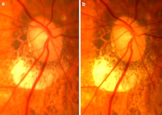
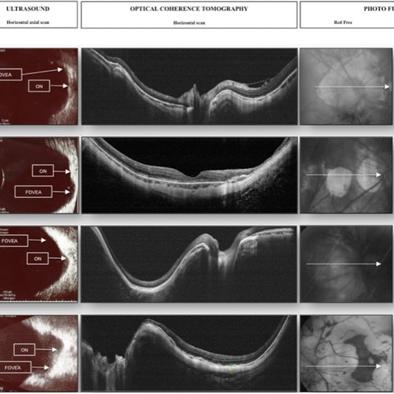
Posterior staphyloma
It is another clinical finding of degenerative myopia. It is abnormal pouching or out pouching of white part of the eye, sclera. As the axial length increases in myopia the sclera also undergo thinning. The abnormal increase in axial length leads to abnormal thinning of sclera. It fails to maintain the structure of curvature. It results in irregular posterior curvature which can be seen in B-scan.
Sub-retinal hemorrhage
The breaks in Bruch’s membrane result in entering of newly formed choroidal vessels in the neurosensory retinal space. These weak vessels rupture and blood leaks out from them leading to hemorrhage.
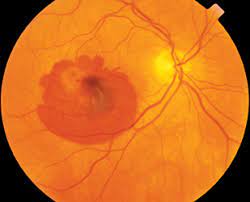
Foster-Fuchs’s pot
These patches occur if the sub-retinal hemorrhage and sub-retinal neovascularization left untreated.
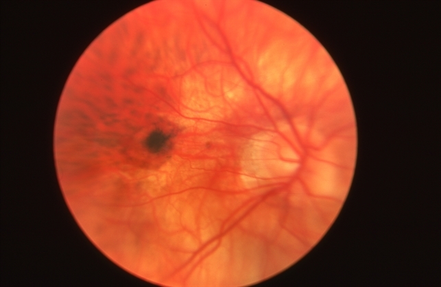
Retinal detachment- invasion of degenerative myopia
As the axial length of the eyeball increases, the posterior vitreous pulls the retinal wall leading to traction.
Treatment for degenerative myopia
Prescription of glasses or contact lenses
These myopes wear high power prescription glasses. Patients also wear contact lenses. The contact lenses enhance more vision field than eyeglasses.
Anti-VEGF therapy
As mentioned above, in degenerative changes in myopia, neovascularization of choroidal vessels occur in sub retinal hemorrhage, these weak and defective blood vessels are of no use. Hence these medicines stop these changes as these can lead to retinal detachment.
Replacement of IOL
The nuclear sclerosis and increase in the refractive index of lens leads to lenticular changes of the lens fibers. This results on cataract. When artificial lens replaces natural lens, it results in better vision.
Vitrectomy
Retinal detachment is a very serious problem that can result in blindness if not treated properly. Vitrectomy is the replacement of liquefied vitreous by gas bubble or oil in the posterior chamber of eyeball. This surgery is very important as retinal detachment can lead to blindness.
Myopia research is very important in the field of ophthalmology. The degenerative changes that occur due to myopia specifically due to degenerative myopia (pathological myopia) can make myopic patients legally blind. In US it is the leading cause of blindness in 2 percent of population.
Founder of EyesMatterMost- an optometry student who loves talking about eyes. I tend to cover topics related to optometry, ophthalmology, eye health, eyecare, eye cosmetics and everything in between. This website is a medium to educate my readers everything related to eyes.