Retinoscopy- everything you must know about it
Retinoscopy is a clinical procedure used to objectively measure the refractive error and accommodative state of the eye—a handheld instrument that projects light into the eye—is used to observe the reflection (or reflex) from the retina to gauge how the light moves within the eye.
The results obtained from retinoscopy allow clinicians to determine the type of refractive error as well.
Types
Static retinoscopy
In static retinoscopy, the patient looks at a distance target, and the accommodation (focus adjustment) of the eye remains relaxed. This is most commonly used to assess refractive errors in adult.
Dynamic retinoscopy
Dynamic retinoscopy involves assessing the refractive state while the patient focuses on a near object. It is mainly used to evaluate accommodation issues or in children to assess near vision.
Principle
The principle behind retinoscopy lies in observing the “retinal reflex,” or the light reflected from the retina when the retinoscope’s light is shone into the eye. By moving the retinoscope and observing how the reflected light moves (with or against the movement of the retinoscope), the practitioner can determine the refractive status.
With movement light reflex
If the light reflex in the pupil moves in the same direction as the movement of the retinoscope, it indicates a “with movement”. This usually signifies that the eye has hyperopia (farsightedness) or low levels of myopia (nearsightedness). In these cases, plus lenses are used to neutralize the movement.
Against movement light reflex
If the light reflex moves in the opposite direction to the movement of the retinoscope, it is known as “against movement”. This typically indicates myopia, or nearsightedness, where the focal point of light is in front of the retina. Minus lenses are used to neutralize this movement.
Attaining neutralization point
The goal of retinoscopy is to achieve neutralization, where no movement of the retinal reflex is observed. The neutral point occurs when the movement of the retinal reflex stops.To reach this neutral point, trial lenses are introduced in front of the patient’s eye, starting with lenses of low power. The practitioner then adjusts the lens power (plus or minus) based on whether a “with” or “against” movement is seen.
Working distance in retinoscopy
One crucial concept in retinoscopy is working distance. This refers to the distance between the practitioner and the patient’s eye during the procedure. Typically, the practitioner performs retinoscopy at a distance of 50-67 cm from the patient’s eye. At this distance, the examiner can easily see the retinal reflex and make accurate assessments of the refractive error. After neutralizing the reflex, the practitioner subtracts the power equivalent to the working distance from the final result.
The examiner uses trial lenses to neutralize the reflex by adding plus or minus lenses until no movement is observed. This is the point of neutrality and corresponds to the patient’s refractive error.
Neutralizing astigmatism
In the case of astigmatism, the retinoscope is used in streak mode to identify the meridians with differing refractive powers. Different lenses are used to neutralize each meridian.
Uses of retinoscopy
- It provides an objective measure of myopia, hyperopia, and astigmatism in the eye of a patient.
- It is especially useful for infants, children, and non-verbal or uncooperative patients who cannot give subjective feedback during a refraction test.
- Retinoscopy can uncover hidden hyperopia, which might not be easily detected through subjective refraction.
- Dynamic retinoscopy evaluates how the eye focuses on a near object, providing insights into the eye’s accommodative function. The examiner observes the movement of the reflex in the pupil. By assessing the direction and speed of the reflex, the clinician can determine if the accommodation is functioning correctly or if there is a lag or lead of accommodation.
Types of Accommodation Disorders Evaluated by Retinoscopy
In patients with accommodative insufficiency, dynamic retinoscopy shows significant lag of accommodation is observed, indicating that the eye is under-focusing
In cases of accommodative excess, the patient over-accommodates for near tasks, resulting in a lead of accommodation during dynamic retinoscopy. The reflex moves “against” the direction of the retinoscope.
In accommodaion infacility disorder is characterized by difficulty in shifting focus between near and distant targets. Dynamic retinoscopy reveals inconsistent reflex movements, as the patient struggles to adjust their focus.
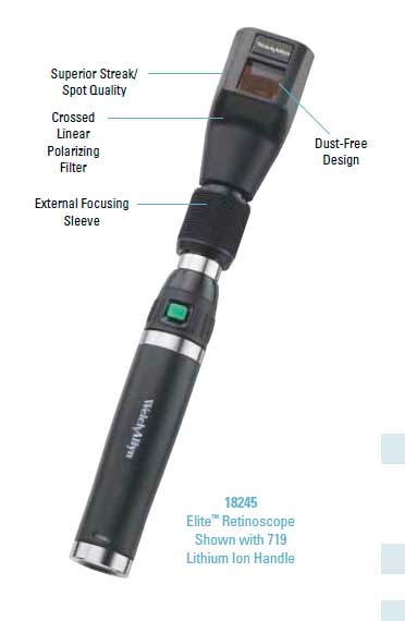
Parts of retinoscope
Illumination system
The retinoscope contains a light source, usually a halogen bulb, that projects light into the patient’s eye.
Mirror
A semi-transparent mirror helps direct light into the eye while also allowing the examiner to see the reflected light from the retina.
Focusing system
The retinoscope has a focusing mechanism that allows the examiner to adjust the direction and brightness of the light beam.
Streak
In streak retinoscopy, a plano-concave lens is used to focus light into a narrow streak, which can be rotated for astigmatism assessment.
Benefits of retinoscope
- Unlike subjective refraction, retinoscopy does not rely on patient responses, making it more reliable test. The more the clinician is expert in handling the retinoscope, the more easy it is to rule out error or accommodation related anomaly with it.
- It is a fast, simple, and non-invasive procedure.
- It is widely used in various clinical settings, from hospitals to school screening programs, due to its effectivenes.
- It is particularly effective in detecting and measuring astigmatism, as it allows practitioners to analyze different meridians of the eye.
- Handheld and easy to carry.
- For patients who cannot provide verbal feedback or those with developmental delays, retinoscopy provides essential information for prescribing corrective lenses.
- Retinoscopy can detect accommodation issues early, preventing long-term vision problems such as eye strain, headaches, and reduced academic or work performance.
Founder of EyesMatterMost- an optometry student who loves talking about eyes. I tend to cover topics related to optometry, ophthalmology, eye health, eyecare, eye cosmetics and everything in between. This website is a medium to educate my readers everything related to eyes.