Optical coherence topography (OCT)- things you need to know
Optical Coherence Tomography (OCT) is a non-invasive imaging technique used to capture detailed images of the retina, the light-sensitive layer at the back of the eye. OCT was first introduced by Dr. David Huang and his colleagues in the early 1990s. The technology was developed at the Massachusetts Institute of Technology (MIT) and quickly became a revolutionary tool in ophthalmology to provide detailed images of the retina.
Working principle of OCT
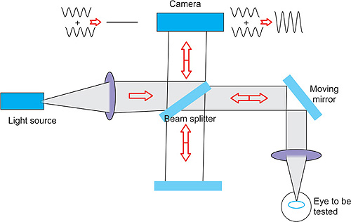
Optical Coherence Tomography (OCT) is based on the principle of low-coherence interferometry, a technique that uses light to capture micrometer-resolution, cross-sectional images of tissues. Here’s a step-by-step breakdown of how OCT works:
OCT uses a broadband light source, typically in the near-infrared range. The light is partially coherent, meaning it has a short coherence length, which is crucial for creating detailed images.
This light is split into two paths:
1- One directed towards the tissue being imaged (the sample arm)
2- The other towards a reference mirror (the reference arm).
The light reflected from the tissue and the reference mirror is combined to create an interference pattern. This pattern occurs only when the light from both paths has traveled nearly the same distance, which allows OCT to measure the depth of structures within the tissue.
The interference pattern varies depending on the optical properties of the tissue, such as refractive index and scattering, which helps in distinguishing between different tissue layers.
The interference signal is analyzed to generate an axial scan (A-scan), representing a one-dimensional depth profile of the tissue at a specific point. This scan measures the intensity of reflected light as a function of depth.
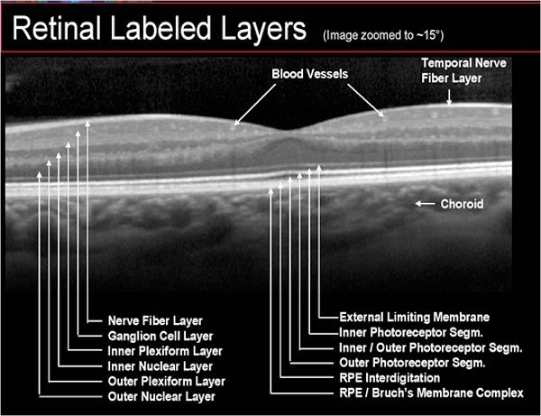
By performing multiple A-scans at different transverse positions, a two-dimensional cross-sectional image (B-scan) is constructed. The B-scan provides a detailed view of the layers of the retina or other tissues.
For three-dimensional imaging, multiple B-scans are taken across a grid, forming a 3D volume of the tissue.
The raw data from the interference patterns are processed using algorithms to enhance contrast and clarity. The result is a high-resolution image that can visualize tissue microstructures, such as retinal layers.
Parts
1. Light Source
It emits a broad spectrum of light that penetrates the eye’s tissues.
2. Interferometer
The interferometer is a critical component that splits the light from the source into two beams: a reference beam and a sample beam.
3. Scanner
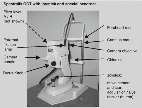
The scanner, often an optical scanner or galvanometer, moves the sample beam across the retina in a precise pattern to capture cross-sectional images (B-scans) of the retina, showing its various layers in detail.
4. Detector
The detector in an OCT system is responsible for capturing the interference pattern created by the combined light beams. This data is then processed to generate a high-resolution image of the retina.
5. Signal Processor
Once the interference pattern is detected, the signal processor converts the raw data into a visual representation of the retina. Advanced algorithms are used to enhance the image quality, making it easier to identify and diagnose any abnormalities in the retinal layers.
6. Display Monitor
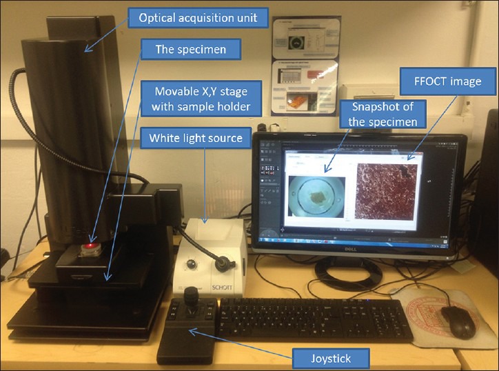
The display monitor shows the final OCT images is used to analyze the condition of the retina.
7. Software Interface
The software interface is an essential part of modern OCT systems. It allows users to control the scanning process, adjust imaging parameters, and store or retrieve patient data. Advanced software also includes tools for measuring retinal thickness, identifying specific layers, and comparing images over time.
Importance
OCT has become an essential tool in the diagnosis and management of various ocular conditions, particularly those affecting the retina.
It also monitors the progression of diseases like macular degeneration, diabetic retinopathy, and glaucoma.
It provides precise images to guide treatments like laser therapy, intravitreal injections, and surgical interventions.
What diseases require this imaging test?
OCT is significant in diagnosing and managing various retinal diseases. It is particularly useful in:
- Age-Related Macular Degeneration (AMD): OCT can detect the presence of drusen, retinal fluid, and other changes associated with AMD.
- Diabetic Retinopathy: It helps in identifying macular edema and retinal thickening, which are common in diabetic patients.
- Glaucoma: OCT measures the thickness of the retinal nerve fiber layer, helping to detect glaucomatous damage.
- Retinal Vein Occlusion: OCT is used to assess macular edema and retinal swelling, which are common in retinal vein occlusion.
Why do they require OCT test?
In several retinal diseases, the thickness of the retinal layers decreases, which can be detected by OCT. These diseases include:
- Glaucoma: Progressive thinning of the retinal nerve fiber layer is a hallmark of glaucoma.
- Retinitis Pigmentosa: This genetic disorder leads to the thinning of the outer retinal layers.
- Advanced Diabetic Retinopathy: Long-standing diabetes can cause thinning of the retinal layers due to ischemia and cell loss.
- Macular Degeneration: In the late stages of AMD, the retinal layers may become thinner due to atrophy.
Side Effects of OCT
OCT is a safe and non-invasive procedure with minimal risks. However, there are a few potential side effects, including:
- Mild Discomfort: Some patients may experience slight discomfort from the bright light used during the scan.
- Eye Fatigue: Prolonged fixation on the scanning target can cause eye fatigue in some patients.
- Rare Complications: Though extremely rare, allergic reactions to the dilating drops used before the OCT scan can occur.
Founder of EyesMatterMost- an optometry student who loves talking about eyes. I tend to cover topics related to optometry, ophthalmology, eye health, eyecare, eye cosmetics and everything in between. This website is a medium to educate my readers everything related to eyes.
